NeuroSPECT wrote:
To answer the original ?. There have been numerous studies using Brain SPECT imaging on autism. SPECT scans show the level of blood flow to the areas of the brain. It has been determined that autistic children have presented consistently with a decrease in blood flow in the temporal area, various degrees of hypoperfusion in the parietal / occipital area and the cerebellum vermis. There has often been an increase of blood flow in the frontal lobes which is consistent with ADD on the hyperactivity end. (if you go to NeuroSPECTofFlorida, there is a full scientific research article on the subject.)
I hope this helps.
NeuroSPECT of Florida
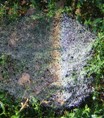
Autism, 16 years old boy. Tc99m HMPAO brain perfusion SPECT demonstrates decreased brain function in the following areas:L Color Blue, < 2 SD Normal Mean), Frontal lobes: Area M, para saggital section of areas 8 and 9 of Brodmann. Area 10. Temporal Lobes: Area 38, anterior Temporal, and 22. There is also hypoperfusion in occipital lobes, Visual association areas, both posterior Parietal areas and cerebellar Vermis and mesial aspects of both cerebellar lobes. Finally there is mild increase of perfusion in Area 24, anterior cyngulate in the left hemisphere. (Color Silver >2SD Normal Mean.[b]
Of course, using SPECT to diagnose brain-related conditions other than epilepsy (and that only at some specific times, not all the time) and a few others is highly questionable scientifically, which is the reason that SPECTs aren't used all over the place to diagnose autism, but only in a fairly small number of places. Worse, I actually saw the SPECT place that I was scanned at, take several different people who were definitively diagnosed with autism and AS and re-diagnose them with something entirely different based only on the SPECT and not on their actual traits. (This includes brain injury, ADD, and drug abuse, among other things. The person accused of drug abuse had never touched brain-altering drugs, legal or illegal, in his life. The person accused of brain injury had no reason at that time for such a severe injury to have happened. There's a reason that particular SPECT center had an awful reputation among parents of disabled children in the area.)
_________________
"In my world it's a place of patterns and feel. In my world it's a haven for what is real. It's my world, nobody can steal it, but people like me, we live in the shadows." -Donna Williams